Germany - Amsterdam Aesthetics originally published at Germany - Amsterdam Aesthetics
Deep learning on annotation-free WSIs for colorectal cancer published to accelerate the development of new high-performance deep-learning algorithms for pathology in the future.

To overcome the obstacle of manual annotation process required intensive labor and time consumption, aetherAI’s deep-learning algorithm detects nodal metastasis of colorectal cancer using its new method of end-to-end training with annotation-free WSIs.
First Published Results of Micrometastasis Detection in Colorectal Cancer
— Joe Yeh, M.D., aetherAI CEO
TAIPEI, TAIWAN, May 25, 2022 /EINPresswire.com/ — aetherAI—Asia’s leading medical image AI solution provider focused on digital pathology and medical imaging AI—has announced the publication of “Identification of nodal micrometastasis in colorectal cancer using deep learning on annotation-free whole-slide images (WSI)” in the peer-reviewed Modern Pathology. At the slide level, the aetherAI algorithm performed well in the identification of both macrometastasis and micrometastasis, with areas under the receiver operating characteristics curve (AUC) of 0.9993 and 0.9956, respectively. This result demonstrates for the first time that micrometastasis can be detected deep learning on whole-slide images without manual annotation. The full article can be accessed online here.
The detection of nodal micrometastasis (tumor size: 0.2–2.0 mm) is challenging for pathologists owing to the small size of metastatic foci. Since micrometastasis lymph nodes are counted as positive nodes, detecting micrometastasis is crucial for accurate pathologic staging of colorectal cancer. Thus, it would be helpful to have an assistive tool for pathologic staging that could detect small metastatic foci in lymph nodes. Although deep learning algorithms have been found to effectively increase the sensitivity and efficiency of micrometastasis detection, the manual annotation process is labor-intensive and time-consuming. To overcome this obstacle, aetherAI has developed a deep-learning algorithm that detects nodal metastasis of colorectal cancer using its new method of end-to-end training with annotation-free WSIs.
aetherAI’s deep learning model uses whole-slide images of regional lymph nodes of colorectal cancer with only a slide-level label (either positive or negative slide). A TAIWANIA 2 supercomputer was used to train a deep-learning model to identify metastasis. At the single-lymph node level, the algorithm performed well at identifying macrometastasis and micrometastasis, with AUCs of 0.9944 and 0.9476, respectively. Visualization using class-activation mapping confirmed that the aetherAI model identified nodal metastasis based on areas of tumor cells.
“The requirement of manual annotation has been a major obstacle to the development of artificial intelligence applications in pathology,” said Joe Yeh, M.D., aetherAI CEO. “Now we have developed for the first time a deep-learning algorithm to detect nodal metastasis in colorectal cancer using undivided, annotation-free WSIs. With no need for time-consuming manual annotation, the aetherAI approach could accelerate the development of new high-performance deep-learning algorithms for pathology in the future.”
The study was conducted in collaboration with pathologists at Chang Gung Memorial Hospital in Taoyuan, Taiwan. The training, validation, and testing sets consisted of 1963, 219, and 1000 slides, respectively.
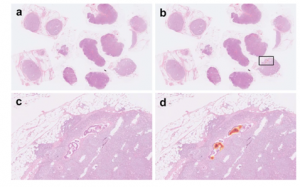
Fig. 1: Example of micrometastasis on aetherAI slide-level testing set.
Using class activation mapping, the original whole-slide image (a) was marked up, with areas of importance for positive prediction highlighted in red (b). A close-up (c) and a highlighted version (d) show that the aetherAI algorithm identified metastasis based on the small areas of tumor cells.
Fig. 2: Examples of micrometastasis in aetherAI single lymph node level testing set.
Using class activation mapping, the original single lymph node images (a, c, e) were marked up, with areas of importance for positive prediction highlighted in red (b, d, f). Note that these areas (asterisked) are small collections of tumor cells (arrowheads).
About aetherAI
Asia’s leading medical image AI solution provider, aetherAI is dedicated to providing cutting-edge solutions for digital pathology transformation, AI-powered diagnostic support systems, and biopharma enterprise services. By leveraging state-of-the-art technologies, aetherAI aims to ease the burden on healthcare professionals and improve diagnostic imaging quality. At the forefront of digital pathology AI, aetherAI partners with top medical centers in the US, Taiwan, and Japan, including UPMC, National Taiwan University Hospital, and Kanazawa University. aetherAI is also a trusted collaborator for such BioPharma companies as Novartis Taiwan and ACT Genomics, and many more are working with us to accelerate AI applications for research efficiency.
Ab Sung
aetherAI CO., LTD.
[email protected]
Visit us on social media:
Facebook
Twitter
LinkedIn

You just read:
EIN Presswire’s priority is source transparency. We do not allow opaque clients, and our editors try to be careful about weeding out false and misleading content.
As a user, if you see something we have missed, please do bring it to our attention. Your help is welcome. EIN Presswire, Everyone’s Internet News Presswire ,
,
tries to define some of the boundaries that are reasonable in today’s world. Please see our
Editorial Guidelines
for more information.
The post aetherAI Marks Deep Learning Advance on Annotation-Free Whole-Slide Images first appeared on Amsterdam Aesthetics.
Germany - Amsterdam Aesthetics originally published at Germany - Amsterdam Aesthetics